A tissue-interface approach for colorectal anastomosis, designed around precise compression and direct submucosal contact.
The product
01
A defined connection at the point of healing.
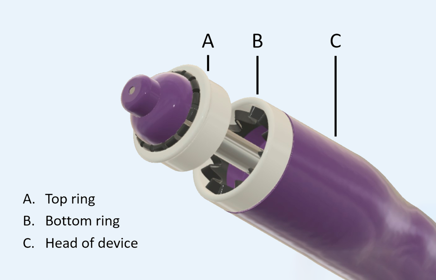
We designed a novel technique based on very strong compression of the bowel with two rings, which tightly fit both bowel ends together. In the surgical procedure, the two intersections of the bowel wall are placed between the rings and brought together.
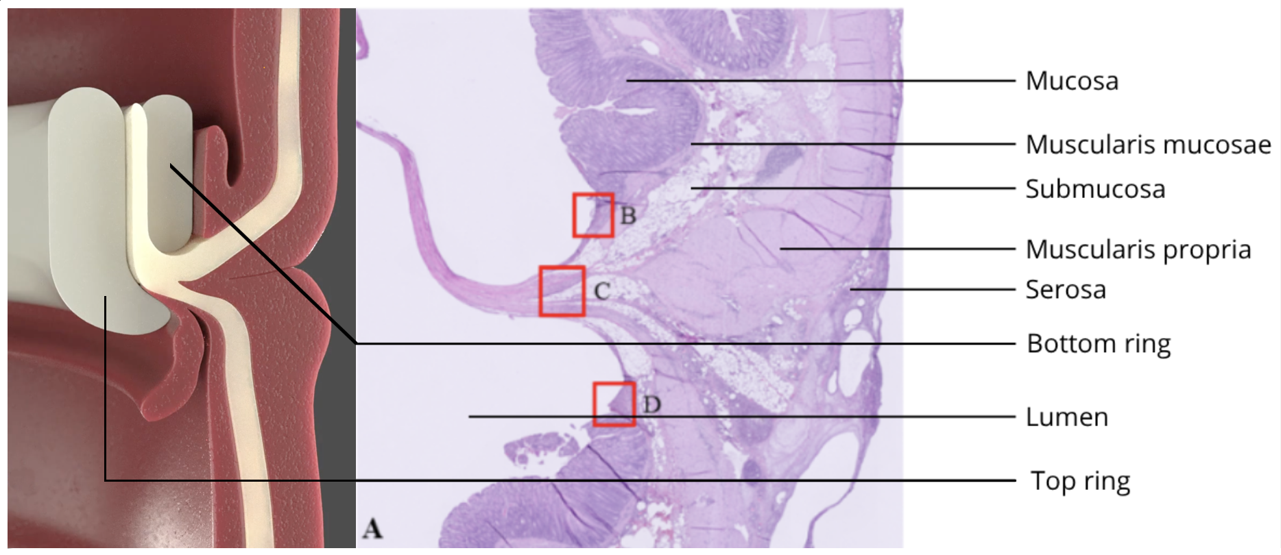
In the compression stage, both bowel walls between the top and bottom ring are heavily compressed following a precise compression profile. It is this precise compression that leads to contact between the submucosal layers of the two intersections of the bowel wall.
The Implican Anastomotic Device has been successfully demonstrated in a surgical procedure on living pigs.
The Implican Anastomotic Device. A: top ring. B: bottom ring. C: head of device.
Implican Technology
The Implican technology aims for a significant reduction of Anastomotic Leakage cases in Colorectal Surgery. By designing our solution as simple as possible, we try to obtain an affordable health care solution which could become widely accessible.
The Implican approach starts with the way the bowel ends meet. It is designed to create a defined contact interface before the biological process of healing unfolds.
Anastomotic healing
A complex process with a precise starting point.
Anastomotic healing involves inflammation, extracellular-matrix remodelling, collagen deposition, angiogenesis and remodelling. Tissue perfusion, tension, microbiota and patient factors also matter.
The Implican hypothesis
Direct tissue contact may support the process.
The submucosa is a collagen-rich layer that contributes to the strength of the bowel wall. The Implican hypothesis is that precise tissue contact gives this complex healing process a better starting point.
Histological healing sequence
02
What was observed preclinically.
Preclinical histology was used to examine the tissue interface over time in porcine specimens.
Preclinical composite showing the Implican compression rings and the histological tissue interface.
POD 1
Tissue apposition.
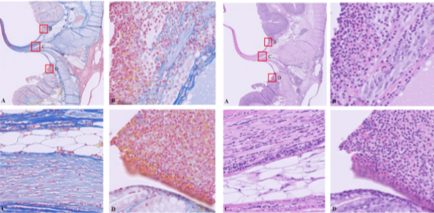
The submucosal tissue is positioned within the Implican compression rings, with an early inflammatory-cell response visible.
MSB and H&E stains from a test animal sacrificed on POD 1. Infiltration of neutrophils is visible in the submucosal layer, mainly at the intraluminal border where the rings stripped off the mucosa and exposed the submucosal surface.
POD 7
Collagen deposition.
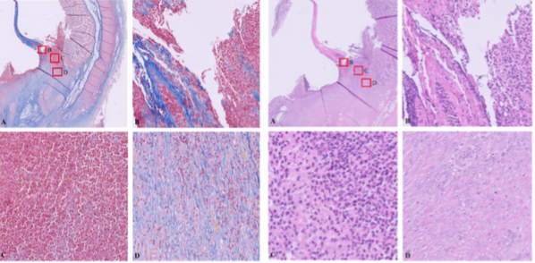
Granulation tissue with fibroblasts and new small blood vessels is present, with significant progression of collagen deposition and fewer neutrophils.
MSB and H&E stains from a test animal sacrificed on POD 7. The MSB staining shows increased fibroblast presence and significant progression of collagen deposition.
POD 30
Remodelling.
In most specimens, luminal ulceration is minimal, the muscularis propria is well aligned, and inflammatory-cell and fibroblast presence is markedly diminished.
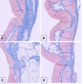
MSB stains from test animals sacrificed on POD 30. The assessed preclinical specimens were consistent with primary wound healing progressing from tissue contact through collagen deposition to remodelling.
Why the absence of staple rows matters
A continuous interface, without staple rows crossing it.
Staples provide mechanical approximation, but they also penetrate and compress bowel-wall tissue. Implican creates an uninterrupted connection without staple rows crossing the healing interface.
The design aims to maintain direct tissue contact while minimising local disruption.
Clinical development
Progressed to initial in-human use.
Clinical development is evaluating technical performance, safety, healing and leakage. Later studies are intended to investigate the impact on temporary stoma use, quality of life and healthcare resources.
Preventing leakage and reducing the need for temporary stomas are development objectives that require prospective validation.